Fluorosis and Renal stones


NOTE: THIS IS AN ONLINE E LOGBOOK TO DISCUSS OUR PATIENT'S DE-IDENTIFIED HEALTH DATA SHARED AFTER TAKING HIS / HER /GUARDIAN'S SIGNED INFORMED CONSENT. HERE WE DISCUSS OUR INDIVIDUAL PATIENT'S PROBLEMS THROUGH A SERIES OF INPUTS FROM THE AVAILABLE GLOBAL ONLINE COMMUNITY OF EXPERTS INTENDING TO SOLVE THOSE CLINICAL PROBLEMS WITH COLLECTIVE CURRENT BEST EVIDENCE-BASED INPUT 

ABSTRACT: Fluorosis a crippling disease is a public health problem with a worldwide distribution and endemic in at least 25 countries with India reporting approximately 25 million people presently affected by fluorosis and 66 million are at risk of developing fluorosis, including children of age 14 years. Here we discuss a case of skeletal fluorosis with a complaint of anuria. We also discuss how fluorosis and how lack of family support crippled this man.

CASE: 

PATIENT HISTORY: The patient was a toddy climber by profession. In the year 2002 patient visited a physician due to colicky pain which was diffuse throughout the anterior abdominal wall, with radiation to his back. The pain was exacerbated by walking and relieved with rest. He was diagnosed with having renal calculi in the right kidney which was operated on and removed. In the year 2007, he fell while climbing a toddy tree and fractured the shaft of his right femur for which ORIF was done. After that he used to walk with support for a few months, then he became normal again but gave up toddy climbing. In the year 2012, he fractured his tibia for which no treatment was done. Later in the year 2012 patient once had a feeling of lightheadedness or dizziness, for which he visited a local physician and was diagnosed with hypertension Since then on regular medication( tab. Ampong 5mg PO/OD) for the same. In the year 2013, he fell in his home and fractured his hip joint for which again no treatment was done. From then onwards he is unable to walk even with support and is bedridden. Gradually, he c/o pain and stiffness in his knee joints which further compelled him to reduce trying to move his joints. In the year 2015 patient developed FIXED FLEXION DEFORMITY grade III of both knees with bilateral knee pain. He started smoking at the age of 25 years and used to smoke 9 bidis/day but stopped 15 years ago due to family pressure and deteriorating health. History of occasional alcohol consumption. He had stopped drinking bore water in 2012 after public awareness program by Govt.
                                 
On 02.02.22 a 65-year-old man visited our hospital in Narketpally, Nalgonda with complaints of anuria since 12pm 01.02.22, with c/o urinary urgency and incomplete voiding of urine, along with the sudden onset of shortness of breath grade 2, paroxysmal nocturnal dyspnea -ve, orthopnea -ve, no chest pain. He complains of decreased appetite in the last few weeks, irregular bowel since last month, a burning sensation since last few days, and abnormal micturition since 1 wk. 

FAMILY HISTORY: It was noted that his wife is also suffering from fixed flexion deformity though with no known history of renal caliculi or some noteworthy problem. We also took into account the fact that his attendant(son) reported that many old people from their community are suffering from fixed flexion deformity.


GENERAL EXAMINATION AT THE TIME OF ADMISSION: The patient is conscious, coherent, cooperative, well oriented to time, place, person. Signs of pallor and pedal edema are present. Mild fever spike. No icterus, cyanosis, clubbing, lymphadenopathy was seen. Both his knees were flexed and at 90 degrees which he was unable to extend, making him unable to move without a wheelchair. Signs of dental enamel molting. His neck was floating midway in the air as if someone kept an imaginary pillow below his head. He is unable to rest his head on the bed and complained of immobility, stiffness, and pain while trying to do any kind of neck movement. The attendant said he has been bedridden like that for the past 10 years.
VITALS :-
TEMPERATURE - 98.6 F
PULSE - 100 BPM
RR - 16 CPM
BP - 160/90 mmHg
spO2 - 98 % @ ROOM AIR
GRBS: 106mg/dl

CVS - S1S2 heard, no murmurs, Apex beat heard over 6th ICS 2cm outside midclavicular line. 
CNS - Intact
RS:- Dyspnea present; BREATH SOUNDS - Vesicular; B/L IAA, ISA crept present
PER ABDOMEN: soft, non-tender 

CHIEF COMPLAINT:  c/o anuria since the day before admission

TREATMENT HISTORY: Tab. Amlong 5 mg PO/OD continuing, Sx for renal calculi, Sx for femur fracture.

TESTS PERFORMED:

ECG report showed little sinus tachycardia. 


Echo showed LV diastolic dysfunction, ?tachycardia
NCCT - KUB(PLAIN): i) Bilateral ureteric calculi causing moderate hydroureteronephrosis
                                    ii) Vesical calculi with diffuse wall thickening
                                    iii) Left renal calculi 4mm size
                                    iv) Minimal bilateral perinephric fat stranding
                                    v) Prostatomegaly
                                    vi) Diffuse bony sclerosis of visualized bones likely suggestive of renal osteodystrophy


RFT : 

          Test                      Result                  Normal Range
  • Urea                  #58mg/dl                  17 - 50
  • Creatinine         #3.0mg/dl                  08 - 1.3
  • Uric acid             6.3mg/dl                  3.5 - 7.2
  • Calcium            #7.8mg/dl                  8.6 - 10.2
  • Phosphorus       2.9mg/dl                   2.5 - 4.5
  • Sodium              144mEq/L                136 - 145
  • Potassium         3.7mEq/L                  3.5 - 5.1
  • Chloride             99mEq/L                   98 - 107
HAEMOGRAM : 
       
          Test                       Result                      Normal Range
  • Hb                         #7.4gm/dl                  13.0 - 17.0
  • TC                           6,900cells/cumm     4000 - 10000
  • Neutrophils              65%                         40 - 80
  • Lymphocytes         #19%                         20 - 40
  • Eosinophils              06%                         01 - 06
  • Monocytes               10%                         02 - 10
  • Basophils                 00%                          0 - 2
  • PCV                       #23.1vol%                 40 - 50
  • MCV                         86.8ft                       83 - 101
  • MCH                         27.8pg                     27 - 32
  • MCHC                       32.0                        31.5 - 34.5
  • RDW-CV                 #15.0%                     11.6 - 14.0
  • RDW-SD                   47.9fl                       39.0 - 46.0
  • RBC count              #2.66millions/cumm   4.5 - 5.5
  • Platelet count            1.79lakhs/cu.mm      1.5 - 4.1
      SMEAR      
          i) RBC - normocytic normochromic
          ii) WBC - within normal limits
          iii) Platelets - Adequate in no. and distribution
          iv) Hemoparasites - No hemoparasites seen
          v) Impression - Normocytic normochromic anemia

URINARY PROTEIN(24 hrs) :
  
            Test                                                          Result                      Normal Range
  • 24 hrs urinary protein                              454.7mg/day                  <150mg/day
  • 24 hrs urine volume                                  1,800ml

COMPLETE URINE EXAMINATION(CUE)

            Test                                                          Result                       Normal Range
  • Colour                                                     Pale Yellow                     Pale Yellow
  • Appearance                                                 Clear                             Clear
  • Reaction                                                      Acidic                             5.0 - 9.0
  • SP Gravity                                                   1.010                          1.001 - 1.035
  • Albumin                                                          +                                 Negative
  • Sugar                                                             Nil                               Negative
  • Bile salts                                                        Nil                               Negative
  • Bile Pigments                                                Nil                                Negative
  • Pus cells                                                      1 - 2                                0-5/HPF
  • Epithelial Cells                                             3 - 4                                0-5/HPF
  • RBCs                                                            2 - 3                               0-2/HPF
  • Crystals                                                          Nil                                  Nil
  • Casts                                                              Nil                                  Nil
  • Amorphous Deposits                                     Absent                              Nil
  • Others                                                            Nil                                  Nil
USG REPORT:
       
      i) B/L gross HUN
      ii) B/L grade II RPD
      iii) Left renal calculi
      iv) Increased texture of kidneys

 CHEST X - RAY REPORT
X RAY REPORT


PROVISIONAL DIAGNOSIS:  AKI secondary to? Acute urinary retention

TREATMENT: 


DAY - 1

      
      IV FLUIDS 2 U NS & 2 U RL @ 100 ML / HR
      INJ LASIX 40 MG IV / BD ( indication - excess fluid buildup due to LVF )
      INJ PAN 40 MG IV / OD
      Nebulization with Duolin and Budecorct 8th hourly( indication - mild URTI )


DAY - 2

S: One session of hemodialysis ( indication of dialysis - anuria ) 

0:  TEMP - Afebrile

BP - 130/90 mmHg

PULSE -86 bpm

RR - 20 CPM SPO2 98% @ RA

CVS -S1S2+, NO MURMURS

RS - BAE +

PA - SOFT, NON TENDER

A:  AKI ON CKD??? WITH LEFT RENAL CALCULUS SIZE 4MM B/L HYDRONEPHROSIS

P:
1) Inj. AUGMENTIN 500mg / IV/BP

2) Inj. LASIX 40mg/IV/BD

3) ORAL FLUIDS ~2L.

4) Inj. PAN 40mg /IV/OD

5) Neb with DUOLIN & Budecort 8th hourly

6) Tab. AMLONG 5mg PO/OD

STRICT I/O CHARTING

DAY 3

S: Urine output increased -1700ml

O: ONE SESSION OF HEMODIALYSIS DONE ON 3/02/2022

TEMP - Afebrile

BP - 130/90 mmHg Pulse 82 BPM

RR - 20 CPM

SPO2 - 98% @ RA

CVS - S1S2+, no murmurs

RS - BAE +

PA - soft, non-tender

A: Acute Kidney Injury?? secondary to Benign Prostatic Hyperplasia? urethral stricture? with left renal calculi size 4mm B/L hydronephrosis

P

1) Inj. Meropenem 500mg / IV/BD

2) Inj. LASIX 40mg/IV/BD

3 ORAL FLUIDS ~2L.

4) Inj. PAN? 40mg /IV/OD

5) Nebulisation with Duolin & Budecort 8th hourly

6) Tab. Amlong 5mg PO/OD

STRICT VO CHARTING


DAY - 4

S: Urineoutput Improved, No fresh Complaints

0: ONE SESSION OF HAEMODIALYSIS WAS YESTERDAY (Total - 2 Dialysis )  


TEMP - AFEBRILE

BP - 130/70 MMHG

PULSE - 96 BPM

CVS -S1S2+, NO MURMURS

RS - BAE +

PA - SOFT, NON TENDER

A: PostRenal AKI Secondary to B/L Ureteric Calculi
     - Moderate Hydrouretero Nephrosis with left Renal Calculi

P :

1) Inj. Meropenem 500mg  IV/BD

2) Inj. LASIX 40mg/IV/BD

3 ORAL FLUIDS ~2L.

4) Inj. PAN 40mg /IV/OD

5) Neb with DUOLIN & Budecort 8th hourly

6) Tab. AMLONG 5mg PO/OD


Strict Input-output charting


DAY - 5

S: Urine output Improved, Patient complaining Of Loss Of Appetite, No Fever Spikes 

0 : Haemodialysis (Total - 2 Dialysis ) .
Output - 1800ml
Input - 1600ml

TEMP - Afibrile
BP - 130/80 mmhg

PULSE - 96 BPM

CVS -S1S2+, no murmurs

RS - BAE +

PA - Soft, non-tender

A: PostRenal Acute kidney injury Secondary to B/L Ureteric Calculi and Bladder Calculi 
     - Moderate Hydrouretero Nephrosis with left Renal Calculi
    - Ossification of the Posterior longitudinal ligament / Diffuse idiopathic skeletal hyperostosis


P:

1) Inj. Meropenem 500mg  IV/BD

2) Inj. LASIX 40mg IV/BD

3 ORAL FLUIDS ~2L

4) Tab. AMLONG 5mg PO/OD


Strict Input-output charting

FINAL DIAGNOSIS: i) Postrenal Acute Kidney injury secondary to B/L ureteric calculi and bladder calculi
ii) Moderate hydroureteronephrosis with left renal calculi. iii) Ossification of the Posterior longitudinal ligament / Diffuse idiopathic skeletal hyperostosis


COMMENT: The patient was recommended Sx to remove renal, ureteric, and bladder stones, but we were not given consent by the patient's attendant. The patient was discharged after treating his chief complaint of anuria.

CASE DISCUSSION :

As we can see this patient is suffering from recurrent renal stones, multiple fractures( indicating weakness of his bones ) from time to time, so in this discussion, we will try to find if there is any correlation between his recurrent renal stones, weak and brittle bones and whether fluorosis can lead to all these problems by gathering and reviewing various articles.

First what lead us to suspect chronic fluorosis?


The patient complains of neck ache, stiffness, rigidity, generalized skeletal discomfort, fixed flexion deformity of his knee joints leading to restricted movement with associated gastric complaints and hails from areas endemic to fluorosis. History of multiple fractures indicating weakness of bones.

Skeletal Fluorosis should be strongly suspected in any person with features of stiffness, rigidity, restricted movements at the spine and joints, bone and joint pains, and who has been residing continuously for >6 months in a fluorosis-endemic area. The clinical investigations for the confirmation of the diagnosis include the following:

  • Haemoglobin%: Anaemia due to reduced erythropoietin activity secondary to fluorosis induced osteosclerosis of medullary cavities (In our patient Hb - 7.4gm/dl, RBC - 2.66 million/cumm)
  • Renal function tests: These may show impaired urea clearance, decreased glomerular filtration rate, increased blood urea nitrogen (In our patient urea - 58 mg/dl)
  • Radiographic features: Increased bony density(osteosclerosis) compact bone thickening, bamboo spine(ankylosing spondylitis), periosteal bone formation. ossification of the attachments of tendons, ligaments, ossification of the post. longitudinal ligament. All of which was seen in our patient.

So, how can chronic fluorosis lead to fractures?

What we learned via reviewing literature...

Prolonged ingestion of fluoride through drinking water, in excess of the daily requirement of 1.5 mg/L as the upper limit (as set by the WHO) is associated with dental and skeletal fluorosis. Signs of skeletal fluorosis become evident on the consumption of 8–10 ppm of fluoride in drinking water for approximately 10 years or more. Our patient is residing in a fluorosis endemic area and had been drinking bore water since birth, only stopping 10 years back.

Vague, diffuse aches, muscle weakness, chronic fatigue, and stiffness of joints with decreased range of motion are common initial symptoms. These symptoms may be dismissed as functional but may, in fact, be early signs of fluoride damage to tendinous insertions and ligaments as well as joint capsules. During later stages, calcification of the bones takes place, osteoporosis in long bones, and symptoms of osteosclerosis where the bones become denser and develop abnormal crystalline structure. In advanced stages, bones and joints become weak rendering movement difficult and painful. Fusion of vertebrae is observed in many areas of the spine. The greatest changes are observed in the spine, particularly in the cervical region. Kyphosis with limited spinal mobility, flexion contracture of lower extremities, and restricted chest wall expansion occur. The “Poker back” spine (kyphosis) is a late manifestation of skeletal fluorosis wherein the entire spine becomes one continuous column of bone. In the final stage, the patient is left crippled. 
The main mechanism of skeletal fluorosis is the fluoride incorporation into the bone hydroxy-apatite, altering the size and structure of its crystals. The fluorapatite formed decreases the mechanical competence of the bone, resulting in abnormal structure and poor quality of bone with an increased risk of fractures.

Diving a bit deeper...

Fluoride can demonstrate biphasic actions (i.e., mitogenic at low concentrations and toxic at higher concentrations).  Fluoride inhibits a unique fluoridesensitive phosphotyrosine phosphatase (PTP) in osteoblasts, which results in a sustained increase in the tyrosine phosphorylation level of the key signaling proteins of the MAPK mitogenic transduction pathway, leading to the potentiation of the bone cell proliferation initiated by growth factors. The fluoride acts in coordination with aluminum to form fluoroaluminate, which activates a pertussis toxinsensitive Gi/o protein on the bone cell membrane, leading to activation of cellular protein tyrosine kinases (PTKs), which in turn leads to increases in the tyrosine phosphorylation of signaling proteins of the MAPK mitogenic signal transduction pathway, ultimately leading to a stimulation of cell proliferation.

Fluoride can act on osteoblasts and osteoclasts in vivo and in vitro. NaF is an anabolic agent capable of increasing bone mass, although the mechanism for its action remains unclear. While NaF may increase bone mass, the newly formed bone appears to lack normal structure and strength. In trabecular bone, fluoride results in an increase in bone volume and trabecular thickness without a concomitant increase in trabecular connectivity. It is this lack of trabecular connectivity that reduces bone quality despite the increase in bone mass. 

Due to a high fluoride concentration in the body, the bone is hardened and thus less elastic, resulting in an increased frequency of fractures. Other symptoms include thickening of the bone structure and accumulation of bone tissue, which both contribute to impaired joint mobility. Ligaments and cartilage can become ossified.

All of these explain how fluorosis leads to his skeletal deformity.

Our next question...Can fluorosis somehow increase the chance of renal stone formation?

What we learned from various articles...

The World Health Organization is acutely aware that fluoride is a kidney toxin, recently citing the finding that approximately 100,000 individuals in the Assam region in India have been taken ill with kidney failure, stiff joints, and anemia as a result of high natural levels of fluoride in the water [WHO 2015]. Similar results have been found in other parts of India [Hindu 2017] and Sri Lanka [Dharmaratne 2015].

 In a recent study to evaluate the role of fluoride in urolithiasis in humans, two areas were selected, a fluoride endemic area (EA) and a fluoride non-endemic area (NEA). The prevalence of urolithiasis was 4.6 times higher in EA than in NEA. Furthermore, the prevalence was almost double in subjects with fluorosis than without fluorosis in the endemic area. No relationship was observed between urolithiasis and the duration of fluorosis. The fluoride levels in drinking water ranged from 3.5 to 4.9 ppm in EA and subjects from this area excreted more fluoride. A comparison of normal subjects (NS) from EA and NEA revealed that endemic subjects tend to have slightly higher mean serum thiobarbituric acid reactive substance (TBAR) levels and excrete more oxalate and fluoride than their non-endemic counterparts. The urinary stone formers (SF) from the two areas showed a similar tendency, though again the difference was not significant. Citrate excretion in SF was almost normal in the EA, but NEA SF had significantly lower excretion levels. Urinary stones from endemic patients had higher fluoride, oxalate, and calcium levels than those from non-endemic patients. In vitro studies suggested that fluoride did not influence the heterogonous mineralization of calcium oxalate. In conclusion, the data suggest that fluoride in vivo may behave as a mild promoter of urinary stone formation by (a) excretion of insoluble calcium fluoride, (b) increasing oxalate excretion, and (c) mildly increasing the oxidative burden.

Among major adverse effects on the kidneys from excessive consumption of fluoride are
immediate effects on the tubular area of the kidneys, inhibiting the tubular reabsorption;
changes in urinary ion excretion by the kidneys
disruption of collagen biosynthesis in the body, causing damage to the kidneys and other organs; and
inhibition of kidney enzymes, affecting the functioning of enzyme pathways.

In another research, 20 urinary stones were taken from patients at a Russian hospital with ion chromatography and conductivity detection. They detected fluoride ions in 80% of the stones, at concentrations of 0.01–4.0mg per gram of stone.
The vast majority of the stones contained very small concentrations of fluorides, below 0.1mg per gram of stone, but two of the stones had fluoride concentrations above 0.5mg. This indicates that the formation of these stones may have been promoted by high concentrations of fluorides in the patients’ urine, perhaps due to the patients ingesting lots of water containing added fluoride or even lots of fluoride toothpaste.
[“In our previous experiment using rats, fluoride was reported to cause renal calcification, whose mechanism was deduced to be due to an increase in parathyroid hormone (PTH) secretion. However fluoride-induced renal calcification that was independent of PTH has not been understood well in the nephron of fluoride-treated animals. Thus, we examined the effect of sodium fluoride on intracellular calcium mobilization in a normal rat kidney epithelial cell line (NRK-52E cells). The calcium accumulation was found to be remarkably increased by the addition of sodium fluoride (NaF). The elevation of [Ca2+]i was demonstrated to be due to calcium entry through nifedipine-sensitive calcium channels.”
SOURCE: Murao H, et al. (2000). Sodium fluoride increases intracellular calcium in rat renal epithelial cell line NRK-52E.Biol Pharm Bull. 23(5):581-4.]

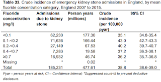
This study shows that there was no association at higher concentrations, and the test of trend indicated a linear trend was not present.
In another investigation, a case-controlled study was conducted on 824 school children aged 8–15 years, where drinking water was contaminated with fluoride. This study showed chronic kidney damage to be higher in the affected children, other than fluorosis, and significantly higher urinary fluoride as compared to the control area children.
Another cross-sectional study conducted on 239 adults (18–77 years old) by monitoring four early kidney injury biomarkers (ALB, Cys-C, KIM-1, and OPN) related to environmental fluoride exposure, indicated possible tubular dysfunction due to fluoride exposure, that might increase susceptibility to the future development of CKD.

After going through these articles we conclude that high fluoride can act as a predisposing factor for renal stones.
CONCLUSION: Our patient emphasizes the importance of considering fluoride as an explanation for osteosclerosis and in diagnosing skeletal fluorosis especially in those who are at high risk from environmental or occupational exposure. Data gathered from different sources do suggest that fluoride may behave as a mild promoter of urinary stone formation by (a) excretion of insoluble calcium fluoride, (b) increasing oxalate excretion, and (c) mildly increasing the oxidative burden, although more extensive research needs to be done to consolidate the fact and understand it's mechanism.
MY EXPERIENCE AS AN ELECTIVE STUDENT: 


Comments

Popular posts from this blog

33 yr old male with c/o pus oozing out from his upp. left third molar region

"MY EXPERIENCES WITH GENERAL CELLULAR AND NEURAL CELLULAR PATHOLOGY IN A CASE BASED BLENDED LEARNING ECOSYSTEM’S CBBLE”

A 60 yr old male aiming to making lifestyle changes rather than take medicine